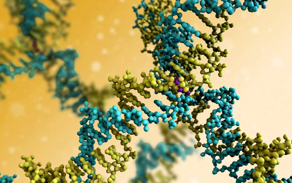
Ever since German anatomist Walther Flemming discovered chromosomes in the mid-1800s, scientists have been actively working to decipher the complex organization of these threadlike structures. In the hundred years following Flemming’s discovery, cell biologists have developed a basic working picture: strands of DNA coil around proteins called histones to form nucleosomes; nucleosomes themselves coil tightly to create chromatin loops; and, finally, chromatin loops wrap around each other to make a full chromosome.
Recent studies now suggest that specific domains exist within the chromatin loops and that the three-dimensional structure and spatial orientation of these domains help to regulate many genomic functions. Several labs around the world have been investigating innovative imaging techniques to gain a more holistic, 3D view of the nucleus and its tangled contents.
Building a Better Fish
One group at the California Institute of Technology (Caltech) in Pasadena is building on a tried-and-true methodology known as fluorescence in situ hybridization (FISH). In this approach, a fluorescent dye is attached to a purified piece of DNA. The tagged DNA is then incubated with the full set of chromosomes from a genome of interest. The fluorescently labeled DNA finds its matching segment on one of the chromosomes and sticks. When researchers look at the chromosomes under a microscope, they can see where the DNA is bound by identifying glowing regions of color.
FISH techniques enable scientists to detect and locate a specific DNA sequence or gene, but they can only look at a few locations in any given sample. The molecular biology lab at Caltech discovered a way to improve FISH results by labeling a single sample repeatedly with many different probes, each with a different color. After the fluorescent probes find and hybridize with complementary DNA or RNA sequences, the tissue sample is imaged and then all of the images are decoded later. The final readout shows thousands of locations up and down the chromosome, resulting in a super-resolved, spatially preserved image of genomic-level processes, including chromatin states and internal and external signaling.
Caltech is still refining the technique, which it calls sequential fluorescence in situ hybridization, or seqFISH, and has co-founded a firm called Spatial Genomics to commercialize it. In late 2021, the team published early results in the journal Science. Using seqFISH techniques, they examined the spatial arrangement of more than 3,000 DNA loci simultaneously in cortex cells taken from a mouse brain. They observed that certain chromosomic features are conserved across cell types and that the 3D spatial arrangements of larger chromatin loops, as well as smaller domains, play a key role in determining the behavior of different cell types.
High-Resolution Results
Other labs are developing similar approaches. The Wyss Institute at Harvard Medical School has introduced a technique known as OligoFISSEQ. FISSEQ, which stands for fluorescence in situ sequencing, came first as a tool to pinpoint the location of thousands of mRNAs simultaneously in intact cells and determine the sequence of bases contained in each of them. By combining Oligopaints — computer-designed probes that attach fluorescent tags to specific DNA sequences — with FISSEQ, researchers can now quickly visualize more regions of the genome, including RNA and the genomic DNA flanking its site of transcription. In a 2020 issue of Nature Methods, scientists explain how they can use OligoFISSEQ techniques to produce 3D maps of 36 genomic targets in four rounds of sequencing. In just five to eight rounds of sequencing, OligoFISSEQ could image thousands of targets.
Another lab at Harvard University is also collecting holistic images of the genome — DNA, RNA and proteins together — using a technique called multiplexed error-robust FISH (MERFISH). MERFISH enables simultaneous imaging of hundreds to more than 1,000 distinct genomic loci at various resolutions in single cells. In the September 2020 issue of Cell, the Harvard team reported that they were able to image approximately 2,200 DNA loci and RNA species in single cells. They also leveraged antibody stains to see additional nuclear structures, including nuclear speckles and nucleoli.
From 2D to 3D
Using these innovative approaches to imaging, cell biologists are gaining a deeper understanding of chromosome structure and function. For decades, a basic 2D model has successfully explained the fundamental mechanics of DNA replication, transcription and translation, but thinking of DNA as a simple string of nucleotide letters only tells part of the story. In reality, a long strand of DNA, along with associated proteins, is folded in a complex manner to form a chromosome. Then, the chromosomes themselves are folded and contorted to fit into a nucleus, much like a group of 20 college students packing themselves into the cab of a Volkswagen Beetle. Scientists are now coming to understand that the complex 3D environment inside a nucleus is just as important to genes expression as the sequence of nucleotides making up those genes.
These innovative techniques, which combine aspects of sequencing and imaging, are finally revealing the spatial organization of the nucleus in ways never thought possible before. And the field is changing rapidly. As Ting Wu, who leads the laboratory at Harvard Medical School that helped to develop OligoFISSEQ, says, “Inventions are happening left and right. I think everyone’s extremely excited to see what the next year’s going to bring.”
Did you enjoy this blog post? Check out our other blog posts as well as related topics on our Webinar page.
QPS is a GLP- and GCP-compliant contract research organization (CRO) delivering the highest grade of discovery, preclinical and clinical drug research development services. Since 1995, it has grown from a tiny bioanalysis shop to a full-service CRO with 1,100+ employees in the U.S., Europe and Asia. Today, QPS offers expanded pharmaceutical contract R&D services with special expertise in neuropharmacology, DMPK, toxicology, bioanalysis, translational medicine and clinical development. An award-winning leader focused on bioanalytics and clinical trials, QPS is known for proven quality standards, technical expertise, a flexible approach to research, client satisfaction and turnkey laboratories and facilities. Through continual enhancements in capacities and resources, QPS stands tall in its commitment to delivering superior quality, skilled performance and trusted service to its valued customers. For more information, visit www.qps.com or email [email protected].


