




As lipid nanoparticle (LNP)-based therapeutics and vaccines continue to gain regulatory approval and clinical adoption, comprehensive evaluation of their immunogenicity has become a critical component of product development. In the fourth and final interview of this series, the QPS Holdings, LLC (DE, USA) team explores the multifaceted immune responses elicited by these products and the application of immunoassays and flow cytometry for characterizing immune responses.
Meet the Subject Matter Experts
 Xun Wang
Executive Director, Bioanalysis
QPS Holdings, LLC
Xun Wang joined QPS in 2009 and is currently serving as an Executive Director of Bioanalysis in the Department of Translational Medicine. In this position, Xun serves as an SME and team manager for developing and validating bioanalytical immunoassays to support biologics drug development, including biosimilars, ADCs and bispecifics. Prior to joining QPS, Xun held Principal Scientist positions at GlaxoSmithKline (PA, USA). Xun received his B.A. in Chemistry from the University of Utah (UT, USA) and his Ph.D. in Biochemistry and Molecular Biophysics from the University of Pennsylvania (PA, USA).
 Monique Putman
Executive Director, Bioanalysis
QPS Holdings, LLC
After completing her degree in Food Technology at Wageningen University (Netherlands), Monique Putman obtained her PhD on the topic of multidrug transporters in Lactococcus lactis. Monique joined QPS Netherlands in 2002 as Senior Scientist. Initially, she supported GLP/GCP method development, validation and sample analysis using HPLC, LC–MS/MS and ligand binding assays. Over time, her focus shifted to biochemical techniques and Monique is currently heading the teams within the department for ligand binding and cell-based assays, acting as subject matter expert.
 Lifeng Tian
Director of Bioanalysis, Flow Cytometry
QPS Holdings, LLC
Lifeng has 15+ years of basic research experience in cell culture, transduction, function assays (cell proliferation, cell migration/cell invasion and animal models), detection assays (Western Blot, IHC/IF, and Co-IP) and molecular skills (PCR, RT-PCR, Microarray, RNA-Seq). She provides high-quality flow cytometry data that phenotypically and functionally evaluates and characterizes the starting materials of apheresis, gene-modified products (CAR-T product) or pre-infusion/post-infusion samples. She is highly skilled at 30-color flow cytometry panel development and troubleshooting, data analysis, interpretation, report writing, presentation and organization. Lifeng has contributed to 10+ publications in Nature, Science, Cells, Nature Medicine, and others.
Meet the Subject Matter Experts
Xun Wang
Executive Director, Bioanalysis
QPS Holdings, LLC
Xun Wang joined QPS in 2009 and is currently serving as an Executive Director of Bioanalysis in the Department of Translational Medicine. In this position, Xun serves as an SME and team manager for developing and validating bioanalytical immunoassays to support biologics drug development, including biosimilars, ADCs and bispecifics. Prior to joining QPS, Xun held Principal Scientist positions at GlaxoSmithKline (PA, USA). Xun received his B.A. in Chemistry from the University of Utah (UT, USA) and his Ph.D. in Biochemistry and Molecular Biophysics from the University of Pennsylvania (PA, USA).
Monique Putman
Executive Director, Bioanalysis
QPS Holdings, LLC
After completing her degree in Food Technology at Wageningen University (Netherlands), Monique Putman obtained her PhD on the topic of multidrug transporters in Lactococcus lactis. Monique joined QPS Netherlands in 2002 as Senior Scientist. Initially, she supported GLP/GCP method development, validation and sample analysis using HPLC, LC–MS/MS and ligand binding assays. Over time, her focus shifted to biochemical techniques and Monique is currently heading the teams within the department for ligand binding and cell-based assays, acting as subject matter expert.
Lifeng Tian
Director of Bioanalysis, Flow Cytometry
QPS Holdings, LLC
Lifeng has 15+ years of basic research experience in cell culture, transduction, function assays (cell proliferation, cell migration/cell invasion and animal models), detection assays (Western Blot, IHC/IF, and Co-IP) and molecular skills (PCR, RT-PCR, Microarray, RNA-Seq). She provides high-quality flow cytometry data that phenotypically and functionally evaluates and characterizes the starting materials of apheresis, gene-modified products (CAR-T product) or pre-infusion/post-infusion samples. She is highly skilled at 30-color flow cytometry panel development and troubleshooting, data analysis, interpretation, report writing, presentation and organization. Lifeng has contributed to 10+ publications in Nature, Science, Cells, Nature Medicine, and others.
Immunogenicity for LNP-based therapeutics and vaccines
In the context of LNP-based therapeutics and vaccines, why is it important to investigate immunogenicity? What are the main immunogenicity assessments that should be assessed and what are the current regulatory recommendations?
Following U.S. Food and Drug Administration (FDA) approvals of several LNP-based therapeutics and vaccines – including Onpattro (2018), Comirnaty (2021), Spikevax (2022) and mRESVIA (2024) – understanding immune responses elicited by LNP-based products has become increasingly important. Both innate and adaptive immune responses can influence safety, efficacy, pharmacokinetics, pharmacodynamics and, where applicable, the feasibility and outcome of repeat dosing. Therefore, comprehensive characterization of product- and payload-associated immunogenicity is an important component of their preclinical and clinical development.
Immunogenicity broadly refers to a substance/therapeutic’s ability to elicit an immune response, which may involve both innate and adaptive immune responses. In the context of LNP-based products, both LNP components and the encapsulated payload, such as mRNA, can be sensed by the innate immune system through pattern-recognition pathways, thereby activating innate immune responses that can modulate the magnitude, quality and durability of subsequent adaptive immune responses. Moreover, when the payload encodes a protein, as in the case of mRNA, the resulting protein may elicit antigen-specific adaptive immune responses, including humoral and cellular immune responses. Overall, the consequences of these immune responses may be beneficial or detrimental depending on the therapeutic indication and the magnitude, quality and durability of the response.
From a regulatory perspective, both the FDA and the International Council for Harmonization (ICH) generally support a risk-based, product-specific approach to evaluating immunogenicity and immunotoxicity rather than requiring a predefined panel of immune assays for all LNP-based products. FDA guidance emphasizes characterizing and mitigating immune responses that may affect the safety, efficacy, pharmacokinetics or pharmacodynamics of therapeutic products, with immunogenicity assessments tailored to product- and patient-specific risks. For therapeutic proteins, the FDA recommends appropriately developed and validated assays for anti-drug antibodies (ADAs), including screening, confirmatory, titration and, where appropriate, neutralizing-antibody assays. ICH S6(R1) provides a framework for the preclinical safety evaluation of biotechnology-derived products, including consideration of immunogenicity and both humoral and cell-mediated immune responses, while ICH S8 provides a weight-of-evidence framework for assessing unintended immunotoxicity effects. Accordingly, for LNP-based therapeutics and vaccines, the selection of innate and adaptive immune endpoints – including cytokines, complement activation, cellular immune responses, antigen-specific antibodies and ADAs – should be scientifically justified based on the product’s composition, mechanism of action, intended indication, and potential impact on clinical safety and efficacy.
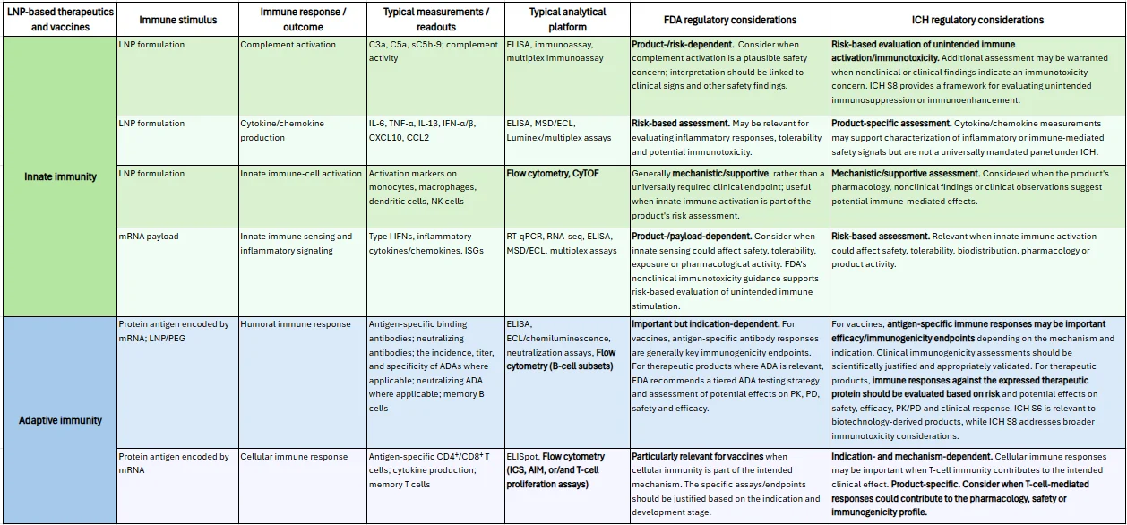
Table 1 provides an overview of immune stimuli, expected immune responses and outcomes, typical measurements and readouts, analytical platforms and regulatory considerations for LNP-based therapeutics and vaccines.
Humoral immunogenicity assessment
Why is it important to investigate the humoral immune response to both the LNP, including PEG-lipid, and the expressed protein, also known as the transgene product?
Investigating humoral responses to both the LNP (especially PEG-lipid) and the expressed/transgene protein is critical because each of these immune responses can arise through different mechanisms and independently affect safety, pharmacokinetics, efficacy and redosing potential.
LNPs containing PEG-lipids can trigger adaptive responses, mainly IgM and IgG-type anti-PEG antibodies, often via T-cell-independent mechanisms due to PEG’s repetitive structure. Pre-existing anti-PEG antibodies are common (roughly 20–70% prevalence, depending on assay and population) from environmental exposure in cosmetics and other products. Treatment-induced or boosted antibodies can accelerate blood clearance of subsequent doses through opsonization and phagocytosis, alter biodistribution, and, in rare cases, lead to hypersensitivity or IgE-mediated anaphylaxis. These effects may reduce cargo delivery efficacy and raise safety concerns, particularly with intravenous or repeated administration.
The expressed transgene protein can elicit classic ADA-like responses similar to protein/antibody therapeutics. Risk depends on sequence homology to endogenous proteins, expression pattern (secreted, membrane-bound or intracellular), dose and route. Antibodies may neutralize activity, clear the protein, form immune complexes, or mediate cytotoxicity (ADCC/CDC) against expressing cells, potentially limiting efficacy or causing tissue damage.
What are the key considerations and typical challenges in developing assays to detect anti-LNP antibodies?
Pre-existing antibodies can elevate baseline signals, reduce dynamic range, and affect interpretation of treatment-boosted responses. Consequently, high prevalence of pre-existing anti-PEG IgM/IgG complicates cut-point determination. Statistical approaches using disease-state or healthy-donor panels could be required; False-positive rates and interpretive thresholds need careful justification, often with confirmatory competition steps (excess free PEG, PEG-lipid, or whole LNP).
Suitable positive controls are difficult to obtain or generate against LNP. The most common and practical positive controls are commercial or custom rabbit or murine antibodies specific to the PEG moiety, particularly the terminal methoxy group of modified PEG. These positive control antibodies are selected because PEG is the dominant surface-exposed immunogenic component of standard LNPs.
Direct labeling of LNP (biotin, SULFO-TAG) for bridging or capture formats is possible but there are several practical issues: stability and integrity risks, epitope alteration and low incorporation efficiency and heterogeneity. Instead, direct coating of unlabeled whole LNP or free PEG provides more success and reproducibility.
Due to the high sensitivity and isotype-independent detection, the bridging assay is the default format for most ADA assays against proteins and peptides. However, this format requires dual labeling, which has proven challenging and less reproducible for LNP/PEG. Therefore, direct ELISA (whole LNP or PEG coated on plates) is the most straightforward format for such assays and can detect both IgM and IgG with generic anti-human detection.
Finally, LNPs as reagents have limited stability (aggregation, lipid exchange, PEG shedding). They require careful storage, lot-to-lot characterization, and often fresh preparation.
What are the similarities, differences and bioanalytical strategies for anti-PEG antibody detection versus anti-LNP antibody detection?
Both detection strategies rely on LBA formats (ELISA, ECL/MSD bridging or direct), multi-tier strategies (screen/confirm/titer), statistical cut points, positive-control reagents, and competition confirmation. Both must address pre-existing reactivity, isotype coverage (IgM/IgG/IgE), and potential clinical impact. Regulatory expectations also overlap; FDA calls for anti-PEG monitoring in PEG-containing products.
Anti-PEG assays typically use free PEG (or PEG conjugates of defined MW, linear/branched, methoxy-terminated) as capture antigen. This provides high specificity for the PEG epitope, good sensitivity with optimized coating/blocking, and easier reagent standardization. Limitations include potential under-representation of the conformational presentation of PEG on the curved LNP surface and lower relevance to particle-level interactions. Sensitivity can reach low ng/mL; multiplex isotype detection (IgM/IgG/IgE) is feasible.
Anti-LNP assays target the full particle surface (PEG-lipid + ionizable lipids + helper lipids + any protein corona or targeting moieties). Formats include whole-LNP coating and labeled intact LNPs in bridging assays. These better reflect authentic epitopes and clinical relevance but face greater challenges with reagent stability, lot variability, non-specific binding and positive-control availability. Sensitivity and specificity may be lower or more variable.
How should anti-expressed protein antibodies be assessed by LBA, and what are best practices for assay format and characterization?
Anti-expressed/transgene protein antibodies should be assessed with a multi-tier LBA strategy similar to ADA assays for protein/antibody therapeutics, adapted for the specific expression context and risk profile.
Platform: Bridging ECL/MSD or ELISA formats using labeled recombinant transgene protein (or relevant domains) for capture and detection are preferred. These are sensitive, relatively isotype-independent, and amenable to drug/target tolerance strategies (acid dissociation, etc.). Direct/indirect formats with immobilized antigen and generic detection are acceptable alternatives, especially early in development, provided both IgM and IgG are covered.
Critical reagents: Use a high-quality recombinant protein matching the expressed sequence (including post-translational modifications if relevant); positive controls (affinity-purified polyclonal or monoclonal anti-protein antibodies); and, where homology to endogenous protein exists, strategies to differentiate treatment-emergent from pre-existing reactivity.
Validation/qualification parameters: Set statistical cut points (screen/confirm/titer) established in relevant matrix (healthy or disease-state). Consider sensitivity (typically ≤100 ng/mL target), selectivity, specificity (competition with unlabeled protein), drug/target tolerance, precision, stability and hook effect. For high-homology proteins, specificity demonstration may be limited.
Immunogenicity assessment by flow cytometry
What innate and adaptive immune responses are commonly assessed for LNP-based therapeutics and vaccines by flow cytometry? When should these assessments be performed during drug development or for specific indications?
Flow cytometry is a well-established, high-throughput, single-cell analytical platform that enables rapid analysis of large numbers of cells and is generally cost-effective for high-throughput cellular phenotyping compared with many other single-cell analytical technologies. These capabilities make flow cytometry well suited for characterizing both innate and adaptive immune responses, as summarized in Table 1. For example, flow cytometry can assess innate immune-cell activation by measuring the expression of activation and costimulatory markers, such as CD69, CD80 and CD86, on dendritic-cell subsets, including plasmacytoid and conventional dendritic cells, as well as on monocytes/macrophages and natural killer cells. For adaptive immunity, flow cytometry can characterize B-cell subsets using markers such as CD19, CD20, CD27, IgD, CD38 and CD138. It is also widely used to evaluate T-cell differentiation (CD45RA, CCR7, CD27, CD28, and CD95), activation (CD69, CD137, CD134, CD25, and HLA-DR), proliferation (Ki-67), cytokine production (IFN-γ, TNF-α, and IL-2), and exhaustion/dysfunction (PD-1, TIM-3, LAG-3, and CTLA-4) across T-cell subsets identified by markers such as CD3, CD4, CD8 and Foxp3. Additionally, flow cytometry can be used to obtain absolute cell counts and/or quantitative measurements based on specific scientific needs. With the increasing adoption of spectral flow cytometry, its high-parameter multiplexing capacity and efficient use of limited samples provide a flexible approach for comprehensively characterizing the complex, multidimensional immune responses to LNP-based therapeutics and vaccines, including changes in immune-cell composition, activation, differentiation and function.
To date, there is no specific regulatory guidance defining when these immune responses should be assessed for LNP-based products. Instead, a risk-based, product-specific approach is recommended. Accordingly, innate immune-cell activation and adaptive immune-cell responses should generally be evaluated throughout development, with the timing and depth of assessment guided by the product’s mechanism of action, formulation, indication, and emerging safety and immunogenicity findings. During early discovery and formulation screening, assessment of innate immune-cell activation can help identify LNP compositions associated with excessive inflammatory responses and support selection of candidate formulations. During preclinical development, in vitro evidence of cellular activation in human cells may provide an important indicator of potential clinical toxicities, even in the absence of corresponding findings in animal studies. These assessments can also provide insights into the product’s pharmacology, tolerability and mechanism of action. During clinical development, immune-cell characteristics should be monitored at time points informed by the product’s pharmacology, nonclinical findings and clinical observations, particularly when immune activation or adaptive immune responses are relevant to mechanism of action, safety, pharmacodynamics or immunogenicity. For vaccines, T-cell responses are generally an important component of immunogenicity assessment, whereas for non-vaccine therapeutics, their evaluation should be driven by the therapeutic mechanism and potential for immune-mediated effects. Overall, innate and adaptive immune-cell responses should be assessed at appropriate stages from preclinical development through clinical studies, using a risk-based strategy tailored to the specific product and indication.
Disclaimer: The opinions expressed in this interview are those of the interviewee and do not necessarily reflect the views of Bioanalysis Zone or Taylor & Francis Group.