



Flow cytometry is a powerful laboratory technique used to analyze the physical and chemical characteristics of cells or particles as they flow in a fluid stream through a beam of light, usually a laser. Flow cytometry offers multiparametric, single-cell, qualitative and/or quantitative data in a rapid and scalable format, making it indispensable for nearly every phase of modern drug development.
What is Flow Cytometry Used For?
Flow cytometers utilize lasers as light sources to produce both scattered and fluorescent light signals that are read by detectors such as photodiodes or photomultiplier tubes. These signals are converted into electronic signals that are analyzed by a computer and written to a standardized format (.fcs) digital data file.
Applications of flow cytometry include PK/PD assessments, cellular phenotyping and functional assays, biomarker detection, proliferation, and toxicology.
- Whether your goal is to explore cell signaling, immune activation, or cell cycle dynamics, we can tailor a flow cytometry method specific to your research needs.
Flow cytometry plays a crucial role in drug development by enabling researchers to analyze individual cells rapidly and with high precision, providing detailed insights into how a drug interacts with specific cell populations.
- In the early stages of drug discovery, flow cytometry is used for target identification and validation by profiling cell populations and detecting differentially expressed proteins that may serve as drug targets.
- It also supports high-throughput screening by allowing thousands of compounds to be tested for biological activity, such as receptor binding, cell proliferation, or activation of signaling pathways. This helps identify potential lead compounds efficiently.
- Essential for studying the mechanism of action of candidate drugs, it enables the analysis of signaling pathways, cell cycle changes, and apoptosis, which helps define how the drug affects cellular processes. Additionally, the technique is widely used in toxicity and safety assessments by detecting off-target effects, measuring cell viability, DNA damage, and immune activation—providing a comprehensive view of potential adverse effects.
- During preclinical and clinical studies, flow cytometry supports pharmacodynamic evaluations by measuring how the drug alters cellular markers and immune profiles in the body. It is particularly valuable in biomarker monitoring and immune profiling, such as tracking T cell subsets or checkpoint inhibitor expression in cancer immunotherapy trials.
- In the development of biologics and cell therapies like CAR-T cells, flow cytometry is used to confirm successful gene modification, characterize immune cell types, and assess their functionality.
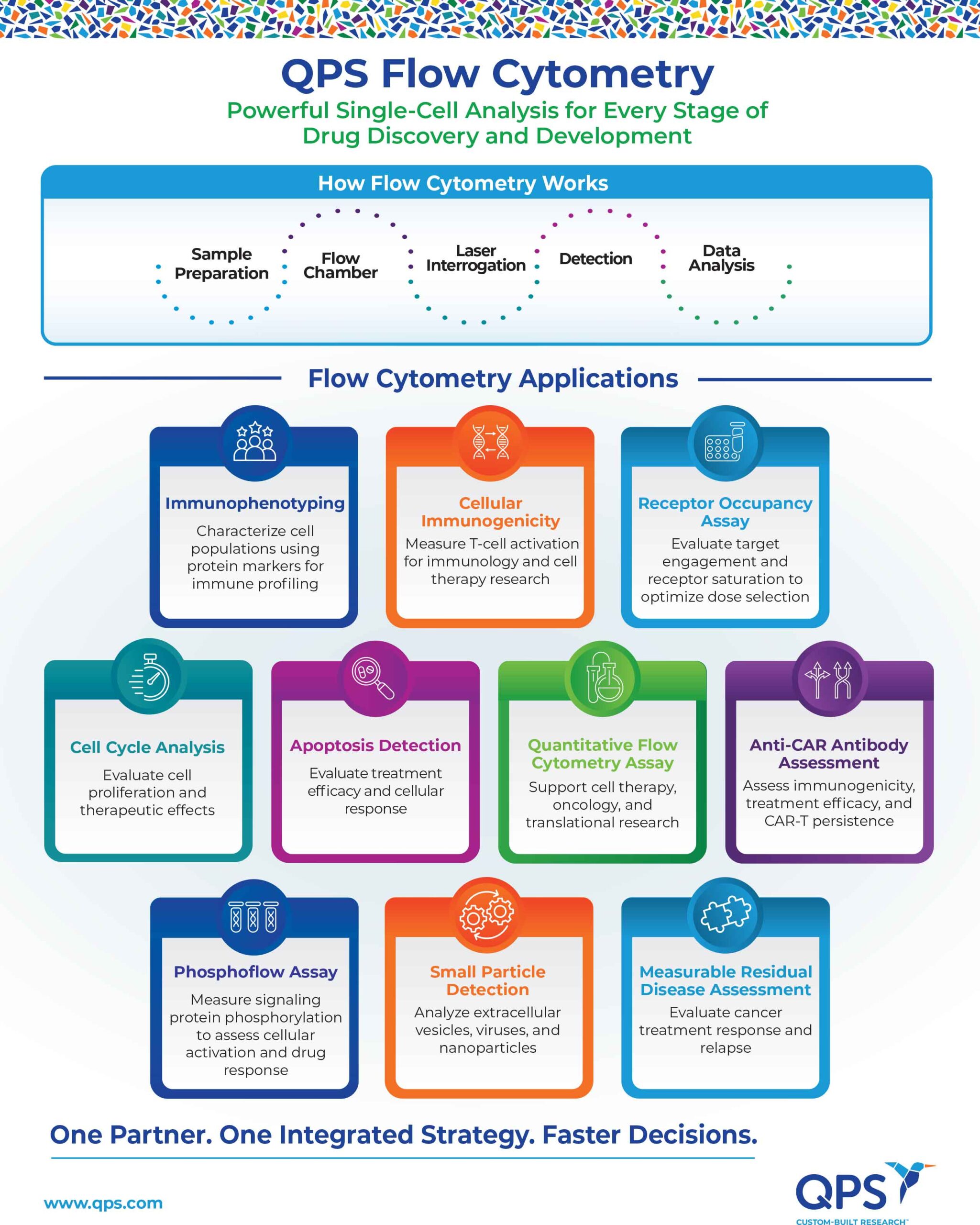
How Flow Cytometry Works
Sample Preparation:
- A suspension of cells (e.g., from blood, bone marrow, or cultured cells) is prepared.
- The cells are often stained with fluorescently labeled antibodies that bind to specific proteins or markers on or inside the cells.
Flow Chamber:
- The sample is injected into a fluid stream that carries the cells single-file through a laser beam.
Laser Interrogation:
- As each cell passes through the laser, it scatters light and any fluorescent markers are excited, emitting light.
Detection:
- Forward scatter (FSC) measures cell size.
- Side scatter (SSC) gives information about internal complexity (e.g., granularity).
- Fluorescence detectors measure light emitted from fluorochromes bound to specific molecules.
Data Analysis:
- A computer collects and analyzes the signals, allowing researchers to quantify and classify cells based on multiple characteristics simultaneously.
What is Flow Cytometry
- Characterizing and classifying cells in a heterogeneous mixture, such as blood, other fluids, or solid tissues. This approach uses the expression of specific proteins, alone or in combination, to categorize cell types based on known functional characteristics.
- Used heavily in immune cell characterization, classification, and leukemia/lymphoma diagnosis.
- Monitoring T-cell function and activation in response to infectious agents, abnormal or malignant cells, vaccine antigens, and other immune stimuli (peptide pools or recombinant proteins).
- Used widely in the development and evaluation of CAR-T and TCR-T cell therapies, infectious disease vaccines designed to elicit T-cell responses, cancer immunotherapies, autoimmune disease treatments, transplantation monitoring, and immunotherapy research.
- Monitoring therapeutic target engagement by quantifying the proportion of cell surface receptors occupied by a drug, enabling assessment of target binding, receptor saturation, pharmacodynamic activity, and optimal dose selection.
- Used widely in the development and evaluation of monoclonal antibodies, bispecific antibodies, CAR-T and other cell therapies, immune checkpoint inhibitors, cytokine- and receptor-targeted therapies, autoimmune disease treatments, oncology therapeutics, inflammatory disease therapies, and clinical pharmacokinetic/pharmacodynamic (PK/PD) studies.
- Monitoring cell cycle progression by quantifying the distribution of cells across the G₀/G₁, S, and G₂/M phases, enabling assessment of cell proliferation, DNA replication, cell cycle arrest, and the effects of drugs or genetic modifications on cell division.
- Used widely in the development and evaluation of anticancer therapeutics, cell cycle inhibitors, targeted therapies, cancer biology research, drug discovery and screening, toxicology studies, stem cell research, regenerative medicine, and studies of cell proliferation, differentiation, and DNA damage responses.
- Monitoring programmed cell death by detecting early and late apoptotic events, membrane integrity, phosphatidylserine externalization, mitochondrial dysfunction, caspase activation, and other markers of cellular response to physiological or therapeutic stimuli.
- Used widely in the development and evaluation of anticancer therapeutics, targeted therapies, immunotherapies, gene and cell therapies, drug discovery and toxicology studies, cancer biology research, neurodegenerative and autoimmune disease research, and investigations of cell survival, cytotoxicity, and mechanisms of cell death.
- Quantifying the expression of cell surface or intracellular biomarkers by measuring the absolute number of target molecules or antigen density per cell, enabling standardized assessment of biomarker expression, receptor density, and cellular phenotype across samples and time points.
- Used widely in the development and evaluation of monoclonal antibodies, CAR-T and other cell therapies, targeted therapies, companion diagnostics, biomarker discovery and validation, immunophenotyping, oncology and immunology research, translational medicine, and clinical pharmacodynamic and diagnostic studies.
- Monitoring humoral immune responses against CAR constructs by detecting anti-CAR antibodies and evaluating their incidence, persistence, and potential neutralizing activity, enabling assessment of immunogenicity, CAR-T cell persistence, therapeutic efficacy, pharmacodynamic effects, and treatment safety.
- Used widely in the development and evaluation of autologous and allogeneic CAR-T cell therapies, CAR-NK and other engineered cell therapies, immunogenicity and bioanalytical studies, clinical pharmacokinetic/pharmacodynamic (PK/PD) assessments, translational medicine, regulatory submissions, and long-term clinical monitoring of therapeutic performance and safety.
- Monitoring intracellular signaling pathway activation by quantifying the phosphorylation status of signaling proteins, enabling assessment of signal transduction, cellular activation, drug response, and pharmacodynamic effects at the single-cell level.
- Used widely in the development and evaluation of targeted therapies, kinase inhibitors, immunotherapies, CAR-T and TCR-T cell therapies, oncology and immunology research, autoimmune and inflammatory disease treatments, drug discovery, biomarker identification, and clinical pharmacodynamic (PD) studies.
- Monitoring and quantifying residual malignant cells that remain after treatment by detecting disease-associated biomarkers at very low levels, enabling assessment of treatment response, disease burden, relapse risk, and therapeutic efficacy at high sensitivity.
- Used widely in the development and evaluation of hematologic cancer therapies, CAR-T and other cell therapies, targeted treatments, immunotherapies, stem cell transplantation strategies, clinical trials, prognostic risk assessment, and longitudinal monitoring of patients with leukemia, lymphoma, and other blood malignancies.
- Detecting and characterizing small biological particles, including extracellular vehicles (EVs), exosomes, microparticles, viruses, and other submicron particles, by analyzing size, concentration, surface markers, and molecular composition at high sensitivity.
- Used widely in the development and evaluation of extracellular vesicle-based biomarkers, liquid biopsy approaches, cell and gene therapies, infectious disease research, immunology studies, oncology applications, drug development, biomarker discovery, and translational medicine.
Flow Cytometry FAQs
Why is flow cytometry considered a powerful analytical technique?
What information can flow cytometry reveal about a cell?
How does flow cytometry support biomarker discovery and validation?
Which therapeutic areas benefit most from flow cytometry?
What makes flow cytometry an important tool for clinical trials?

Contact Us
Would you like to learn more about other QPS capabilities?
Visit: https://www.qps.com/
Set Up a Meeting: https://www.qps.com/contact
Email: info@qps.com